- 28 September 2026
- Lina Kocienė, Vilnius University information
Lithuanian Scientists Develop a Non-destructive Methodology to Analyse Tooth Enamel Microcracks
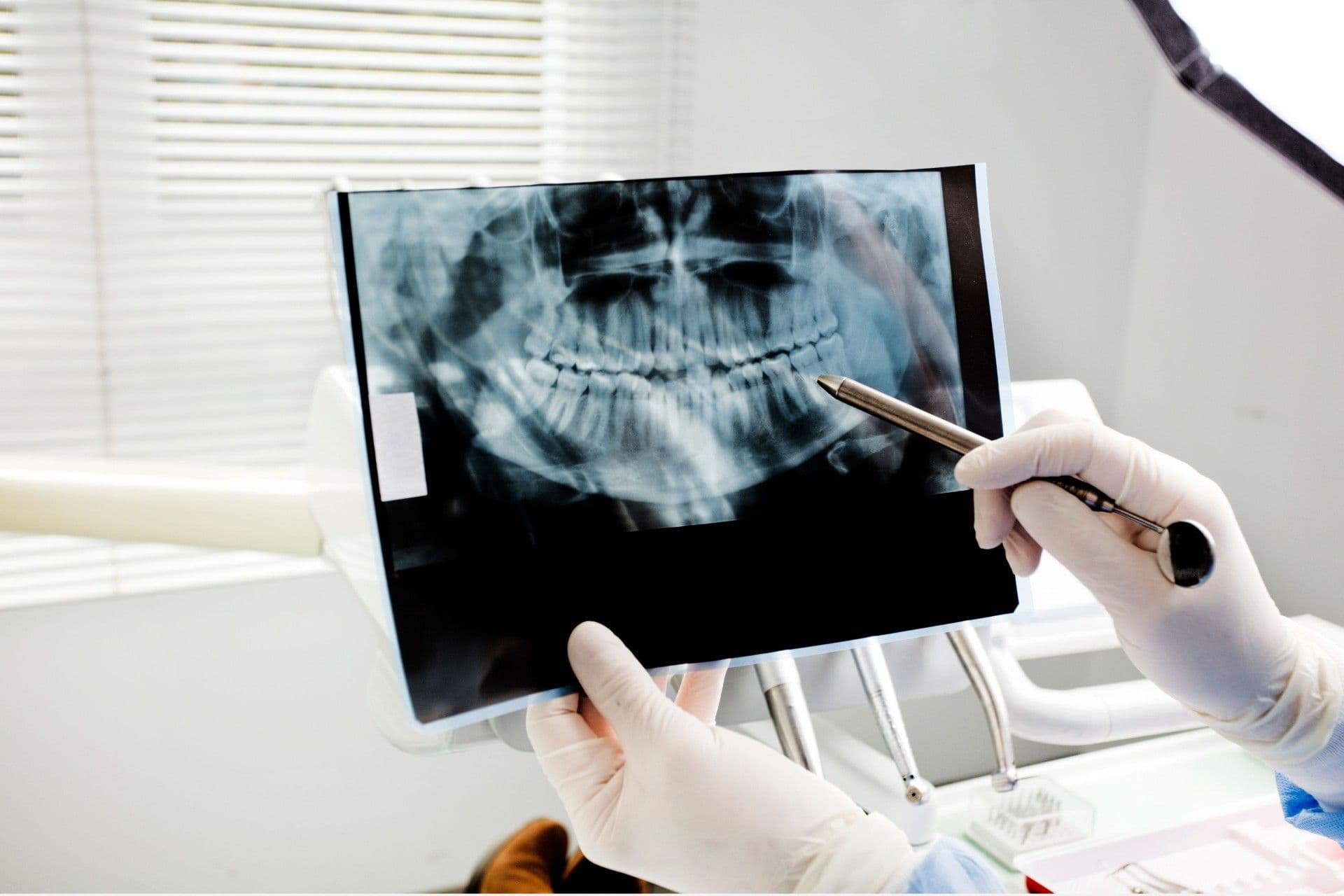
Researchers at Vilnius University (VU), together with partners from Kaunas University of Technology, the Foundation for Research and Technology – Hellas (FORTH), and Swinburne University of Technology in Australia, have developed a new non-destructive methodology that enables the integration of data from measurement modalities based on different principles to provide a more accurate description of microcracks in tooth enamel within intact (uncut) teeth. The results of the research have been published in the international scientific journal 'Measurement'.
The researchers have developed a spatial data fusion (registration) methodology which, using a 3D mesh corresponding to the tooth surface, combines data from two methods – X-ray micro-computed tomography and photoluminescence spectroscopy. Additionally, energy-dispersive X-ray spectroscopy is used to assess changes in the elemental composition of the enamel. In this way, data about a specific location on the tooth obtained using different instruments are integrated, allowing for a comparison between crack-affected and pristine enamel.
'The essence of the study was not merely to examine a microcrack, but to understand what is happening at the crack-affected site. Different measurement methods provide different information: one enables the structure to be visualised in 3D, another reveals the optical response, and a third shows the elemental changes. By combining this information, we can describe the damage much more accurately,' noted Dr Irma Dumbrytė, senior researcher from the Institute of Dentistry at the VU Faculty of Medicine, who led the research.
Cracked tooth enamel is revealed by both its structure and optical response
The study analysed human premolars with enamel microcracks visible to the naked eye. The cracks were further confirmed using scanning electron microscopy. Photoluminescence spectrum measurements were then taken in both the cracked and pristine areas and the data obtained were combined with 3D tomography information.
The results showed that there was a statistically significant difference between the photoluminescence spectra of cracked and pristine enamel. This means that, in future, the optical response could be used as an additional indicator for identifying areas of enamel damage, and such information could be incorporated into clinical practice, particularly where an image of the tooth structure alone is insufficient to provide all the necessary information.
The study also revealed that the elemental composition of the enamel changes at the sites of microcracks. Analysis showed an increased carbon content – the average value at the crack sites was 42% higher than for pristine enamel. The carbon-to-oxygen ratio also increased – from 0.46 in the pristine areas to 0.84 in the cracked areas. The calcium and phosphorus contents changed comparatively little between the cracked and pristine areas.
These results illustrate how the damage to the enamel structure, its elemental composition and its optical response are all related. Such comprehensive characterisation may be important for gaining a better understanding of microcrack formation, their progression and their potential impact on the integrity of the tooth structure.
One methodology for different hard materials
One of the most important findings of the study is that the spatial data fusion methodology that was developed can be applied beyond the analysis of tooth surfaces. The 3D surface mesh-assisted registration framework proposed by the researchers allows data from measurement modalities to be integrated, even when their resolution, measurement principles and coordinate systems differ. According to the researchers, this opens up possibilities for applying the methodology to other hard materials where a single investigative method is insufficient to characterise surface defects.
'Most importantly, we have not merely developed an approach specifically for microcracks in teeth, but a general principle for reliably combining information from different measurement modalities. This could be important both for further dental research and for the broader characterisation of defects in materials,' Dr Dumbrytė noted.
From laboratory research towards future diagnostics
The researchers emphasise that the current study has demonstrated a methodology, not a tool for clinical diagnosis. Nevertheless, the results obtained lay the groundwork for further research.
According to the researchers, in the future, higher-resolution X-ray imaging technologies, such as synchrotron nanotomography, could facilitate the research of even finer cracks and a more detailed analysis of their geometry. There are also plans to integrate Raman microspectroscopy data into the same approach, which would provide additional information about the chemical composition of the material.
It is expected that by combining various research methodologies, it will be possible in the future to develop new non-invasive or minimally invasive methods for assessing the condition of enamel, specifically designed for the diagnosis of microcracks in clinical practice.
The study was carried out as part of the project, “Multi-Spectral Volumetric Teeth Microcracks Investigation”, funded by the Research Council of Lithuania (contract No. S-ITP-25-6, principal investigator Dr Irma Dumbrytė). Some of the measurements were carried out at FORTH in Heraklion (Greece), utilising the NFFA-Europe Pilot international access activity funded by the European Union’s Horizon 2020 programme (grant agreement No. 101007417, proposal ID-572).